
Diffusion-weighted imaging (DWI) and tractography (DTI)
Diffusion-weighted imaging (DWI) and tractography (DTI) play a crucial role in understanding brain function, as it relies on a complex network of nerve fibers that connect different regions of the brain. DTI, a specialized form of diffusion tensor imaging, utilizes the movement of water molecules along these nerve pathways to visualize these structures. This imaging technique not only reveals the intricate bundles of nerves but also aids in characterizing and diagnosing white matter disorders in conditions such as tumors, stroke, and epilepsy. Furthermore, it provides valuable information about the location and proximity of important nerve fibers in relation to tumors, enhancing the precision and accuracy of surgical interventions.
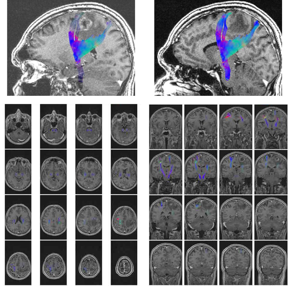
The above provided images depict the results of DTI imaging analysis, specifically the reconstruction of motor fiber pathways.
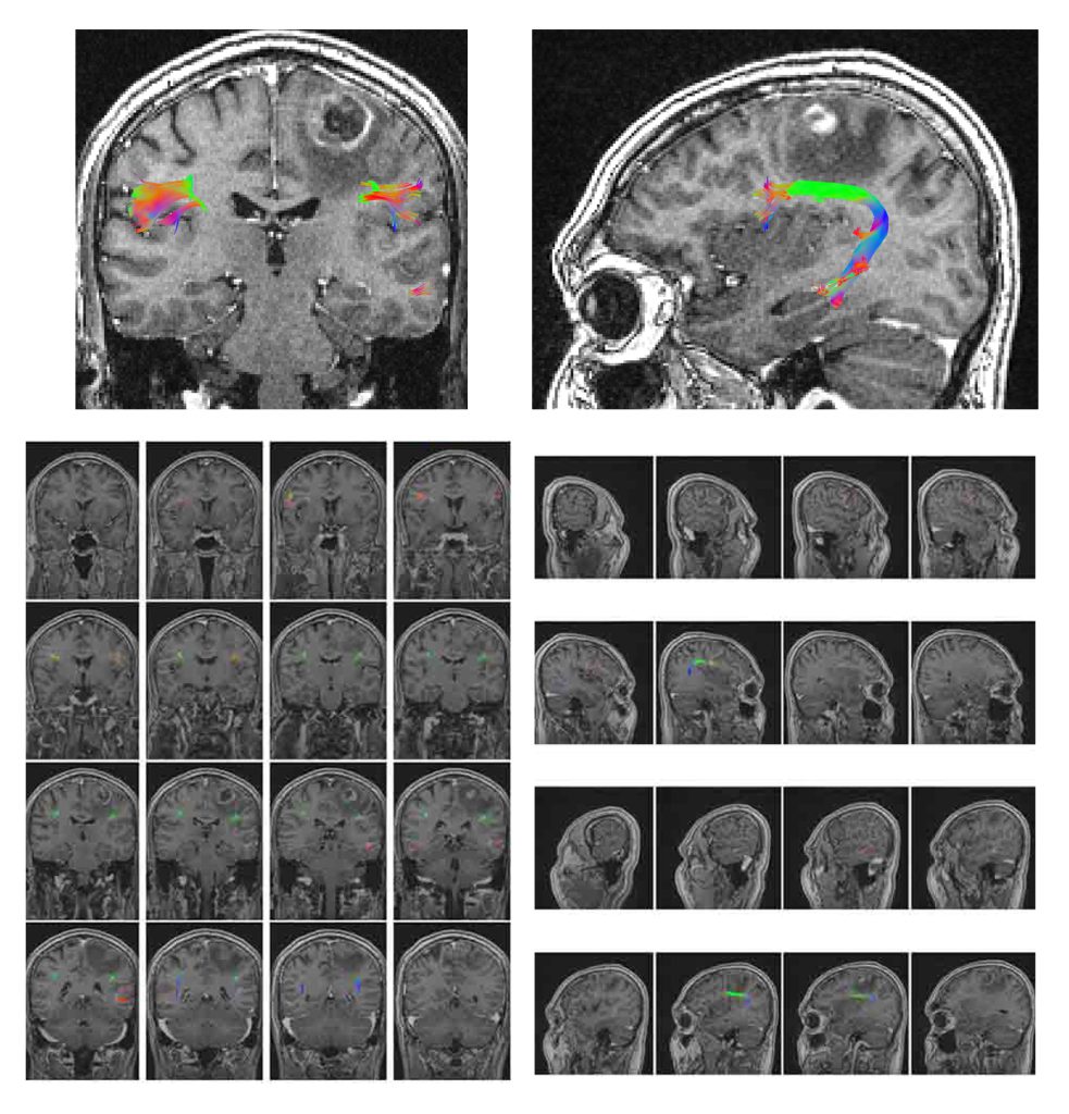
The above provided image showcases the reconstruction of speech pathway fibers.
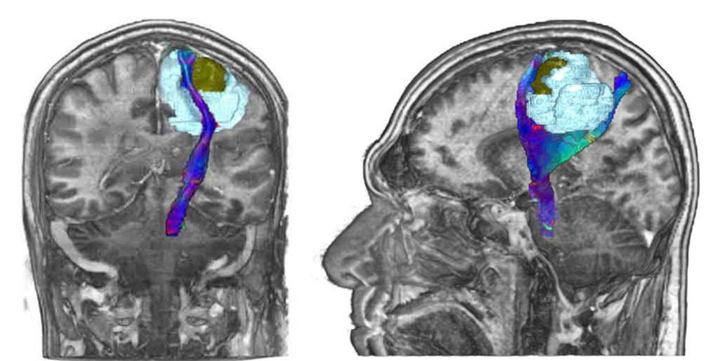
The accompanying image displays a 3D reconstruction of a tumor (highlighted in yellow), edema (indicated in blue), and motor fibers located in close proximity to the lesion.
Neural pathways that can be analyzed are as follows
- Motor pathways, such as the corticospinal fibers, which are responsible for coordinating voluntary movements.
- Language pathways, including the Arcuate fibers, IFOF (inferior fronto-occipital fasciculus), Uncinate fasciculus, SLF (superior longitudinal fasciculus), and ILF (inferior longitudinal fasciculus), which are involved in speech production and comprehension.
- Memory pathways, such as the fornix fibers, which play a role in memory formation and retrieval.
- Visual pathways, which comprise the optic nerve, optic tract, and optic radiation, and are responsible for transmitting visual information from the eyes to the brain.
- Olfactory pathways, involving olfactory nerve fibers, which enable the perception of smell.
- Brainstem fibers, like the MLF (Medial Longitudinal Fasciculus), are involved in coordinating eye movements and maintaining balance.
- Cerebellar pathways, such as cerebellar connections, contribute to motor control, coordination, and balance.
For which clinical scenarios is DTI imaging typically requested? DTI درخواست ميشود
- Presurgical planning prior to brain surgery.
- Planning before invasive brain treatments, such as radiotherapy.
- Diagnosis of brain and nerve diseases that have impacted the structure of brain fibers.
- Specialized or differential diagnoses that cannot be achieved with conventional imaging methods.
Standard prescription guidelines include
- Specify the specific type of fiber or tract and the precise location of the brain imaging areas required, or indicate the type of structural imaging necessary in the prescription.
- Clearly state the reason for requesting DTI imaging in the prescription.
- Provide a comprehensive medical history of the patient, including relevant therapeutic interventions, in the prescription or accompanying forms available to the physician.