Seizure Mapping
Seizure mapping plays a crucial role in the diagnosis and management of epilepsy and other seizure disorders. Utilizing MRI technology, we are able to visualize the intricate structure of the brain. Through careful analysis of these images, we can pinpoint the precise areas implicated in seizure activity. By conducting accurate mapping of these regions, we acquire invaluable insights into the origins and pathways of seizures, ultimately guiding us towards more effective treatment strategies.
At NIAG Clinic, we employ cutting-edge MRI machines to capture high-resolution images of tumors. Our team of experts, including experienced radiologists, utilize advanced software and specialized techniques to meticulously analyze these images. Through this comprehensive analysis, we are able to identify and map the seizure focus as well as its associated networks.
The benefits of utilizing MRI for seizure mapping are substantial. Through precise identification of the areas implicated in seizure activity, we are able to facilitate treatment decision-making, surgical planning, and the implementation of targeted therapies. Our comprehensive approach guarantees that patients receive individualized care that is specifically tailored to their distinct needs and seizure patterns.
Seizure Mapping, a comprehensive multi-protocol brain imaging approach, involves obtaining various images and maps of the entire brain from three different directions: coronal, axial, and sagittal. This includes standard brain protocols as well as high-resolution structural imaging in all three directions. Additionally, T1 and T2-FLAIR imaging is performed in at least two directions, along with SWI for improved visualization. If a lesion is present, contrast-enhanced brain imaging is conducted. Furthermore, in order to assess the possibility of microstructural changes and FCD, DTI or VBM techniques are employed. Volumetry is utilized to evaluate atrophy, while CSI and MRS are employed to investigate underlying metabolic diseases and growth disorders.
Specific indications for prescribing this service include
These indications are based on evidence-based paraclinical and clinical criteria. The service is recommended in cases where the presence of epilepsy-related factors and the need for precise assessment and intervention have been established. The number of cases in which this service is indicated for an individual patient will vary depending on their specific condition and medical history.
- Diagnosis of underlying pathological factors, structural abnormalities, and physiological changes that contribute to the development of epilepsy.
- Identification and characterization of brain lesions responsible for epileptic activity, including determining their type and extent.
- Presurgical planning and preparation for invasive procedures aimed at treating epilepsy.
Standard prescription guidelines include
- Specifying the type and location of brain imaging areas required or the specific type of structural imaging in the prescription.
- Clearly stating the rationale for requesting specialized imaging in the prescription.
- Provide a comprehensive medical history of the patient, including relevant therapeutic interventions, in the prescription or accompanying forms available to the physician.
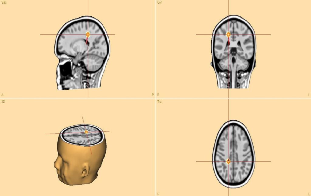
The Seizure Mapping protocol includes an analysis conducted in a patient to identify the epileptic focus. (This analysis is performed to pinpoint the specific area or region in the brain that is responsible for generating epileptic activity).