
Tumor mapping
Tumor mapping plays a crucial role in the diagnosis, treatment planning, and monitoring of various tumor types. Through the utilization of perfusion imaging, we can assess the tumor's blood flow patterns, offering valuable insights into its vascularization and potential invasiveness. Furthermore, MRS imaging enables us to analyze the tumor's chemical composition, aiding in the differentiation of various tumor types and providing crucial information regarding their metabolic activity.
At NIAG Clinic, we employ cutting-edge MRI machines to capture high-resolution images of tumors. Subsequently, our team of expert radiologists, along with the aid of advanced software and sophisticated algorithms, meticulously analyze these images to generate precise maps of the tumor and its adjacent structures.
Tumor mapping using MRI offers numerous advantages. Through precise characterization of tumor blood flow and metabolic activity, we can effectively guide treatment decisions, evaluate response to therapy, and monitor the progression of the disease. Our comprehensive approach ensures that patients receive individualized and customized care, ultimately enhancing their outcomes and overall quality of life.
In general, Tumor Mapping, a multi-protocol brain imaging technique, encompasses various protocols that involve both with and without brain injection, FLAIR sequence, DWI/ADC, and either MRS or Perfusion methods.
Specific indications for prescribing this service include
(Accurate indications for prescribing this service include evidence-based paraclinical and clinical criteria and also the number of cases in which the provision of this service in a patient has an indication).
- differential diagnosis of tumors from other brain and nerve diseases,
- treatment evaluation for brain surgery, brain radiotherapy, and chemotherapy,
- determination of the type, characteristics, and grading of brain-nerve tumors,
- design prior to brain surgery,
- planning before invasive brain treatments such as radiotherapy (Presurgical Planning).
Standard prescription guidelines include
- Specifying the type and location of brain imaging areas required or the specific type of structural imaging in the prescription.
- Clearly stating the rationale for requesting specialized imaging in the prescription.
- Provide a comprehensive medical history of the patient, including relevant therapeutic interventions, in the prescription or accompanying forms available to the physician.
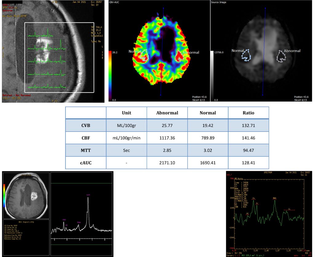
The analysis was carried out for the Tumor Mapping protocol in a single patient.